Why silicone oil?

The higher effective atomic number (Z) of silicone oil compared to that of water, or water equivalent fluids, results in increased attenuation (compared to water) of low energy radiation which traverses the oil. This occurs because attenuation for the low energies emitted by radionuclides such as I-125 and Pd-103 is predominantly the result of photoelectric interactions.

In photoelectric interactions the photon is absorbed and an electron is released. This electron loses its energy very rapidly, so the photon's energy is all deposited in matter very close to the site of the photoelectric interaction. There is no scatter. In general, the probability of photoelectric interactions (attenuation coefficient value) is proportional to Z3. The conditions that increase the probability of photoelectric interactions are low photon energies and high-atomic-number materials.

The objective of replacing the vitreous humor with silicone oil during eye plaque brachytherapy is to reduce the radiation dose to ocular regions outside of the tumor volume by surrounding the tumor with a fluid that has a higher effective atomic number than normal vitreous humor. The oil, however, can only attenuate radiation that traverses it. Therefore, destinations that are in the path of primary radiation which only passes through regions such as sclera, tumor, lens or anterior chamber will receive essentially the same dose regardless of what fluid occupies the vitreous chamber.

The ratio of radiation attenuation in silicone oil to attenuation in normal vitreous fluid will be proportional to the distance traversed in fluid in the vitreous chamber. The path length through fluid that crosses the vitreous chamber between a radiation source and a point of dosimetric interest will vary with the location of the dosimetry point, the tumor location, size and shape, and the position of the radiation source in the plaque.

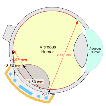
The diagram on the right illustrates a standard COMS style plaque with a Silastic™ silicone seed carrier. Primary radiation emitted from the sources closest to a point of interest such as the fovea, if not removed by collimation, will deliver the dominant contribution to the dose at that point.

For this particular plaque and tumor location, primary radiation from one of these nearby seeds travels 8.36 mm, first through the seed carrier and then through sclera to reach the fovea. This radiation never encounters the vitreous chamber. Radiation from the furthest seed, which delivers the least dose at the fovea owing to the approximately inverse-square relationship of radiation intensity as a function of distance from the source, first travels 11.3 mm through seed carrier, sclera and tumor before reaching vitreous fluid. Only the final 4.95 mm of its total 16.25 mm journey passes through vitreous fluid where it would encounter silicone oil.
RadiationPath

Radiation emitted from this same peripheral seed, which is directed towards the ora on the far side of the eye, first traverses 2.39 mm of seed carrier and sclera, followed by 20.48 mm through fluid as it crosses the vitreous chamber. The original intensity of this radiation, by the time it reaches the far side of the eye, will be primarily reduced by the following factors; inverse-square geometry (.0018), attenuation and scatter in water (.737), and a pair of additonal attenuation correction factors to account for greater attenuation in both the silicone seed carrier (.82) and silicone oil (.51) in the vitreous in comparison with otherwise water equivalent material. By far, the most significant factor affecting the dose received on the far side of the eye is inverse-square.


Silicone Oil Attenuation
SiliconeOil

When calculating the attenuation of radiation as it passes through the eye, the vitreous humor is, by default, assumed to be water equivalent. If the vitreous will be replaced by silicone oil, enable this option to correct for the greater linear attenuation coefficient of silicone oil compared to water. The correction is performed by ray tracing the primary radiation path through a 3D nonhomogeneous voxel model the eye with resolution of 0.2 mm. Voxels within the eye are classified as sclera, tumor, lens, aqueous humor and vitreous humor. In the examples that follow, in order to expressly look at the effects of vitreous oil, the attenuation of all tissue voxels except those representing the vitreous humor are considered to be water equivalent.

VoxelModel

The linear attenuation coefficient for silicone oil is found in the Dose Constants tab of each radionuclide's physics file and may need to be customized for whichever silicone oil variant you are using. Applicable only to gamma sources. Disabled by default.


Semi-posterior large tumor

When the vitreous is replaced by silicone oil, the greatest proportional dose reduction compared to water will occur at locations where the radiation travels the longest distance through the oil. Depending upon the shape and height of the tumor, and assuming the plaque lies directly below the tumor base, this will occur at or near the diametrically opposite side of the eye from the plaque. Radiation as it passes through tumor and other nominally water equivalent materials will be attenuated at the rate of water, so the magnitude of dose reduction compared to water equivalent vitreous will be inversely related to the volume of the tumor. For example, large tumors will displace more oil than small tumors, resulting in less opportunity for attenuation in oil compared to small tumors.

MeridianPlanes

For ocular regions close to the plaque rim, the radiation passes almost entirely through only sclera or tumor, so there is no opportunity for attenuation in oil. Dose reduction, compared to a water equivalent environment, increases with distance from the upper surface of the tumor, reaching its maximum effect on the diametrically opposite side of the eye from the tumor and plaque.

RetinalDiagrams

These retinal-dose-area histograms compare a silicone oil filled vitreous chamber (solid lines) with water equivalent vitreous (dashed lines) for otherwise identical semi-posterior plaque treatments. For a 24 mm diameter eye, 8 mm tumor height, EP model 1821 3rd generation plaque, and posterior location studied in this example, dose to the tumor base (brown curves) and a surrounding 2 mm margin (green curves) is identical. Dose to the optic disc (magenta) is slightly reduced and dose to fovea (beige) and macular (orange) regions is reduced by a few Gy.

Histograms

COMS vs EP Conformal treatment, Semi-posterior large tumor

This example compares treatments using a 16 mm COMS plaque with silicone oil in the vitreous chamber vs an EP model 1821 3rd generation plaque using conformal treatment planning and water equivalent vitreous.

COMSMeridians

For a critical ocular region close to the plaque rim, such as the optic disc in this example, the radiation reaching the disc passes almost entirely through only sclera or tumor, so there is little opportunity for attenuation in oil. The most effective way to reduce dose to a critical structure near the plaque is conformal treatment planning. For example, deleting the seed closest to the disc and redistributing its strength amongst the remaining seeds, combined with individual seed collimation can significantly reduce the dose to the disc without compromising base or margin coverage.

COMSDiagrams

These retinal-dose-area histograms compare a silicone oil filled vitreous chamber (solid lines) with water equivalent vitreous (dashed lines) for this semi-posterior case using different plaques and treatment planning strategies. The eye is 24 mm diameter, the tumor height 8 mm tumor, the plaques are a 16 mm COMS plaque with silicone oil in the vitreous vs an EP model 1821 3rd generation plaque using conformal treatment planning and water equivalent vitreous. These histograms demonstrate that both plaques deliver 100% Rx dose coverage of both the tumor base (brown curves) and a surrounding 2 mm margin (green curves). The conformal plan, however, delivers a maximum scleral dose that is about 100 Gy less, and maximum disc dose (magenta curves) that is about 25 Gy less than the COMS+oil plan. The mean dose to the fovea, about 32 Gy, is nearly identical for both plaques.

COMSHistograms

Semi-anterior large tumor

The dose reduction of silicone oil compared to a water equivalent vitreous will probably be, proportionally speaking, the greatest for ciliary tumors and other anterior tumor locations where the optic nerve and macular region of the eye are as far as possible from the tumor. In this example we look at an 8 mm tall anterior tumor.

MeridianPlanesAnterior

RetinalDiagramsAnterior

These retinal-dose-area histograms compare a silicone oil filled vitreous chamber (solid lines) with water equivalent vitreous (dashed lines) for otherwise identical anterior plaque treatments. For a 24 mm diameter eye, 8 mm tumor height, EP model 1821 plaque, and anterior location studied in this example, dose to the tumor base (brown curves) and a surrounding 2 mm margin (green curves) is identical. Dose to the optic disc (magenta) is reduced from about 16 Gy to 10 Gy (by about 37%), and dose to the fovea (beige) is reduced from about 23 Gy to 16 Gy (by about 30%). The percentage dose reduction is proportionally significant but the absolute dose is low and probably below radiation tissue tolerance thresholds. The absolute dose will be much lower for shorter anterior tumors for which the Rx point will be further from the macula as illustrated in the next example.

HistogramsAnterior

Semi-anterior small tumor

In this example we look at a much shorter, 4 mm tall semi-anterior tumor. Note: the Rx height was simply set to the tumor apex, no attempt was made to assure base margin coverage in this example.

MeridianPlanesSmallAnterior

RetinalDiagramsSmallAnterior

These retinal-dose-area histograms compare silicone oil filled vitreous chamber (solid lines) with water equivalent vitreous (dashed lines) for otherwise identical anterior plaque treatments. For a 24 mm diameter eye, 4 mm tumor height, EP model 1821 plaque, and anterior location studied in this example, dose to the tumor base (brown curves) and a surrounding 2 mm margin (green curves) is identical. Dose to the optic disc (magenta) is reduced from about 7.5 Gy to 4.5 Gy, and dose to the fovea (beige) is reduced from about 10 Gy to 7 Gy. The percentage dose reduction is proportionally significant but the absolute dose is small, probably well below tissue tolerance dose thresholds.

HistogramsSmallAnterior

Ciliary and Iris tumors
CiliarySetup

Dose reduction to the macula and optic disc will be, proportionally, the greatest for ciliary and iris tumor treatments where the plaque is as far as possible from the macula and therefore radiation reaching the macula must first pass through as much silicone oil as possible.

In this example we look at the model EP1930 corneal dome plaque which is designed for full iris and ciliary coverage. This calculation assumes a 24 mm diameter, slightly oblate eye, 1 mm tumor height, an EP model 1930 cornea dome plaque centered on the anterior pole, and a Rx of 85 Gy to the entire iris and ciliary region.


CiliaryMeridians

CiliaryDiagrams

These retinal-dose-area histograms compare a silicone oil filled vitreous chamber (solid lines) with water equivalent vitreous (dashed lines) for otherwise identical, full ciliary, treatment. Dose to the tumor base (brown curves) and a surrounding 2 mm margin (green curves) is identical. Dose to the optic disc (magenta curves), macula (orange curves) and fovea (beige curves) is reduced by about 43% from 6 Gy for water equivalent vitreous to 3.4 Gy when silicone oil replaces the vitreous. The percentage dose reduction is proportionally significant but the absolute dose is small.

CiliaryHistograms