RESULTS
The Macintosh implementation (16MHz 68020 processor) requires approximately 3 secs per seed to calculate a 40 X 40 dose matrix with lip detection activated, and is about 20% faster when lip detection is disabled. Interpolating the dose matrix and generating the screen display requires from 3 to 30 secs, depending only on the size of the display window and the type of display surface requested (planar or 3D perspective projection). The time required to generate a screen display is independent of the dose matrix resolution. Although matrices up to 160 X 160 points are available at the cost of proportionately longer computation time, no visual improvement in isodoses within the hypothetical tumor volume was apparent between the 40 X 40 and 80 X 80 modes. The higher resolution modes which generated the figures in this report are primarily intended to reduce aliasing in the penumbral region near the lip. A typical plaque with 10 seeds normally requires < 60 sec to calculate and display a dosimetry matrix adequate for clinical purposes, and about 1 sec to generate a dose table for the central axis and critical structures.
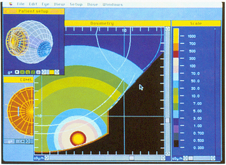
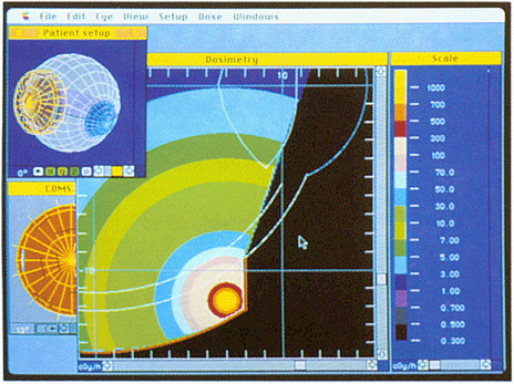
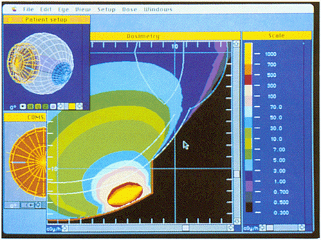
The partial visibility algorithm was tested using a 16 mm diameter COMS plaque. A single 4 mCi seed of I-125 was assigned to a slot near the center of the plaque (Fig. 7a), near the lip (Fig. 7b), and to a radially oriented slot near the lip of a modified plaque (Fig. 7c). The displayed isodose distributions are consistent with the expected shielding effects. Shielding of the sclera adjacent to the plaque is greatest for the seed near the lip and nonexistent for the centrally located seed. Some aliasing in the penumbral region is evident for the radially oriented seed, but is of little practical concern. This aliasing results from the limit of five iterations imposed (to improve speed) on the pvr calculation.
(a)
(b)
|
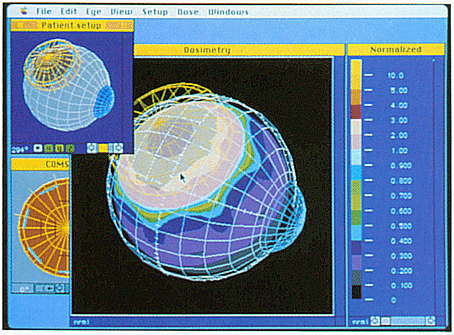
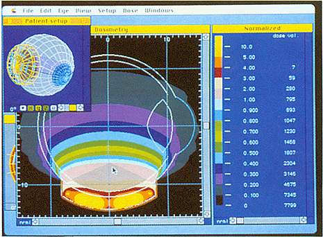
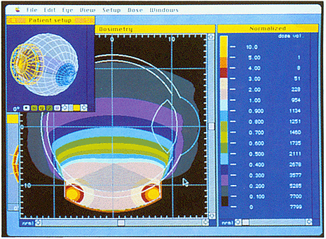
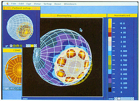
Fig. 8. Comparison of a peripheral loading pattern versus a uniform loading pattern for the COMS 16 mm diameter plaque. The dose distribution is calculated for the plane z = 0. The display has been normalized to a value of 1.0 at 6 mm from the plaque surface on the central axis of the plaque, near the apex of a hypothetical tumor. The nominal position of the retina is indicated by the inner white semi-circle, inset 1 mm from the scleral surface. The tumor is roughly conical in shape, and is indicated by the darkened region within the eye. Dose volume is calculated in mm³. (a) 13 equal sources filling all available slots. (b) 7 equal sources in the peripheral ring.
|
Alternative loading patterns for the plaque were studied to model pre-treatment planning capability, again using the 16 mm COMS plaque loaded with I-125 seeds. The plaque was centered on the y-axis at the intersection of the equator and lateral (90°) meridian (Fig. 4). Dose matrices were calculated in the plane z = 0 which transects both the plaque and eye. In the first case, 13 sources of equal activity filled all of the available slots in the plaque (Fig. 8a). This was compared to seven sources of equal activity in the peripheral ring of slots (Fig. 8b). The dose distributions of Figure 8 have been normalized to a value of 1.0 at 6 mm above the surface of the plaque near the apex of a hypothetical 5 mm tumor. While dose to the tumor volume appears to be similar, retinal dose appears to be more homogenous for the peripheral loading pattern. A dose-volume histogram for I mm³ voxels within the eye was calculated for each case. The results indicate that the peripheral loading pattern increased the volume of eye tissue receiving a dose >= 4X the normalization dose by only I mm³ while decreasing the volume >= 2X by 52 mm³. For this particular geometry, the improved homogeneity of the peripheral loading pattern was achieved at the cost of a slight increase in dose to the lens and to the sclera near the peripheral edge of the plaque.
(a)
(b)
|
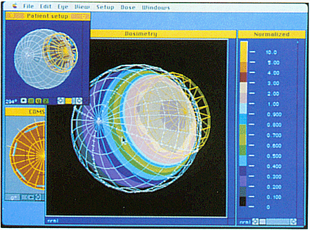
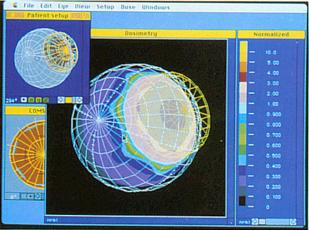
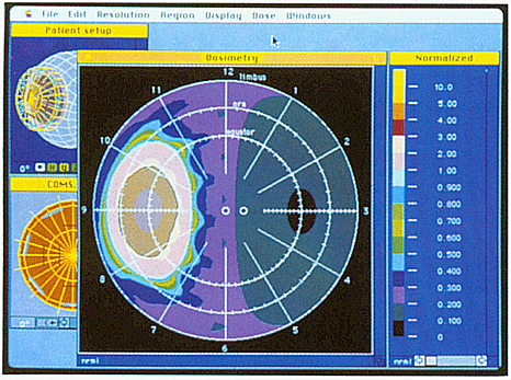
Fig. 9. Dose distributions on the scleral and retinal surfaces for the COMS 16 mm plaque with 7 equal I-125 sources in the peripheral ring. The display has been normalized to a value of 1.0 at 6 mm from the plaque surface on the central axis of the plaque. (a) 3-dimensional perspective projection view of the scleral dose distribution. (b) Funduscopic diagram of dose to the retina.
|
The dose distribution on the scleral surface and the retina (assumed to be 1 mm inset from the scleral surface) are modeled in Figures 9 & 10 for the plaque depicted in Figure 8b. All dosimetry is normalized as in Figure 8. The scleral dose distribution is plotted as a 3D perspective projection in Figure 9a. The retinal dose distribution is plotted in the manner of a funduscopic diagram in 9b. Localized areas of high dose on the sclera adjacent to each seed, which were not readily apparent in Fig. 8b are now clearly seen in Fig. 9a. Outside the lip perimeter indicated by the yellow wire-frame in Figure 9a, a distinctive lobular pattern is observed in the 0.1 to 0.7 relative dose range. This pattern apparently results from partial visibility of alternating sources. Figures 10a & 10b display views of the retinal dose from anterior and posterior vantage points. Figure 10c is identical to 10b except that lip collimation is ignored. The dose distribution in Figure 10c suggests that the lip, rather than anisotropy is the source of the lobular pattern. For this particular geometry and orientation, in which the plaque is adjacent to the optic nerve, the calculated dose to the center of the optic disc in Figure 10c is roughly twice that of 10b.
Abstract |
Introduction & Methods |
Discussion & References